Molecubes Modular Benchtop Preclinical SPECT Imaging System
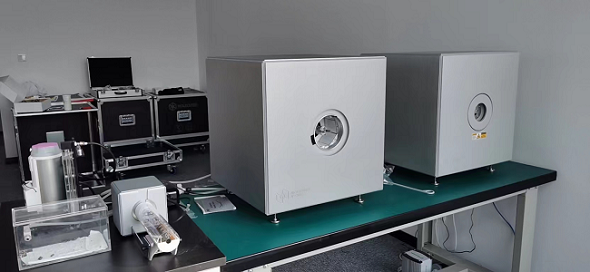
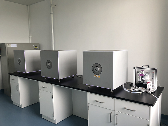
The γ-CUBE is our high-sensitivity, high-resolution SPECT imager for whole body mouse and rat imaging.
Patented lofthole technology and laser sintered collimators, combined with high-resolution detectors, result in a high-end true benchtop imager.
Image reconstruction software, developed in-house, guarantees fast imaging and excellent image quality.
All common SPECT-labeled therapeutic and diagnostic imaging tracers can be imaged.
Intuitive, wireless acquisition software, combined with our multimodal small-animal bed, enables easy and modular multimodal imaging when used along with the X-CUBE (CT) and β-CUBE (PET).
Detail
Product Principles
Single-Photon Emission Computed Tomography (SPECT)
The basic principle of small animal SPECT is: First of all, the organism needs to ingestion a radioisotope imaging agent with an appropriate half-life. After the imaging agent reaches the fault location that needs to be imaged, gamma photons are emitted from the fault due to radioactive decay. The gamma camera located in the outer layer detects gamma photons through the collimator, and the detected high-energy gamma rays are converted into a low energy but large number of light signals through the scintillator. Through the photomultiplier tube, the optical signal is converted into electrical signal and amplified, and then converted into digital by analog/digital converter, and input to the computer for processing. You can get a three-dimensional picture of how the developer accumulates in the organism.

Brief Introduction
The γ-CUBE is our high-sensitivity, high-resolution SPECT imager for whole body mouse and rat imaging.
Patented lofthole technology and laser sintered collimators, combined with high-resolution detectors, result in a high-end true benchtop imager. Image reconstruction software, developed in-house, guarantees fast imaging and excellent image quality. All common SPECT-labeled therapeutic and diagnostic imaging tracers can be imaged. Intuitive, wireless acquisition software, combined with our multimodal small-animal bed, enables easy and modular multimodal imaging when used along with the X-CUBE (CT) and β-CUBE (PET).


Product Characteristics
Highest Throughput – Fast and sensitive/low dose
High-end Performance – PET/CT and SPECT/CT without compromise
Compact – light and mobile
Ease of use – functional and straight-forward
Best Uptime in preclinical industry
Fast workflow
Thanks to our fast and simple CUBEFLOW and intuitive user interfaces, 3D images are obtained using a turnkeysolution.
High precision
Extremely high-quality imaging in terms of resolution and precision and field of view at the smallest footprint.
Modular
Our CUBES are designed to work bothas stand-alone units and in modular combinations so you can optimize yourworkflow and increase throughput.
Benchtop
MOLECUBES offers exquisite whole body mouse and rat imaging while fitting on top of any classic lab bench.
Best uptime
98,6% System uptime and a dedicated support team with extensive preclinical imaging expertise.
User friendly
Developed by end users, we pride ourselves in our intuitive software that allows even the most novice users tocapture beautiful images.

Application Fields
1. Oncology research: tumor genesis, metabolism, apoptosis, lymphatic vessel imaging, hypoxia imaging, tumor receptor research, tumor gene imaging, tumor treatment evaluation, etc.;
2. Research on neurological diseases: brain perfusion imaging, brain receptor imaging, brain metabolism imaging, neurodegenerative diseases (Alzheimer's disease, Parkinson's disease), inflammatory molecular imaging, etc.;
3, cardiovascular system disease research: heart disease (coronary heart disease, heart failure, heart transplant evaluation), atherosclerosis, thrombosis, cardiac nerve receptor research, etc.;
4. Gene therapy imaging: molecular imaging evaluation of gene therapy;
5, cell tracer technology: stem cell therapy, all kinds of diseases (nervous system diseases, myocardial infarction, etc.) after cell therapy evaluation;
6, new drug research and development: preclinical experiments (biological distribution, pharmacokinetics), drug target confirmation, drug lead compound screening;
7. Others: bone disease research, infectious disease research, metabolic disease research, etc.
Application
1、SPECT imaging of nervous system
Brain SPECT imaging in rats: 99mTc-HMPAO: cerebral blood perfusion imaging agent for the diagnosis of cerebrovascular diseases, brain trauma, epilepsy, dementia, and brain death; It is used to study the brain function of mental illness and normal brain physiological function. 99mTc-HMPAO, which enters the brain tissue, changes its configuration and becomes a water-soluble compound, which cannot pass the blood-brain barrier again and remain in the cell, so it can remain in the brain for a long time. 120 MBq, 30 min uptake, 30 min SPECT.

2、Cardiovascular SPECT imaging
Myocardial SPECT imaging in mice: the drug 99mTc-MIBI reflected the myocardial blood perfusion volume at the time of injection, 63.27MBq (1.71mCi) @start acquisition, and the acquisition time was 40min.
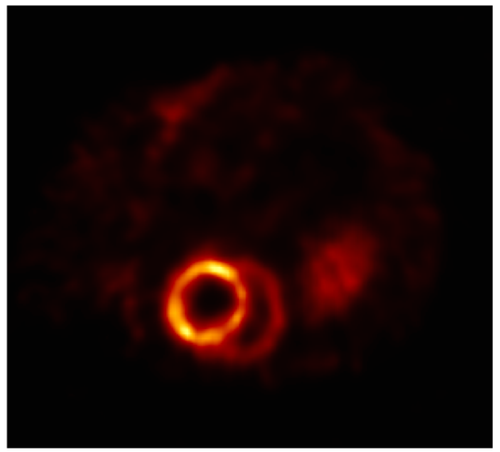
The cardiac blood flow was observed before and after stroke and after medication in rat model of cardiovascular stroke.

3、Bone SPECT imaging
SPECT/CT combination images, bone imaging of mice: 99mTc-HDP, 94.35MBq (2.55mCi) @start acquisition, acquisition time of 60min.

SPECT/CT combined imaging of bone in rats: 99mTc-HDP, 200MBq, acquisition time 45min.

4、Renal SPECT imaging
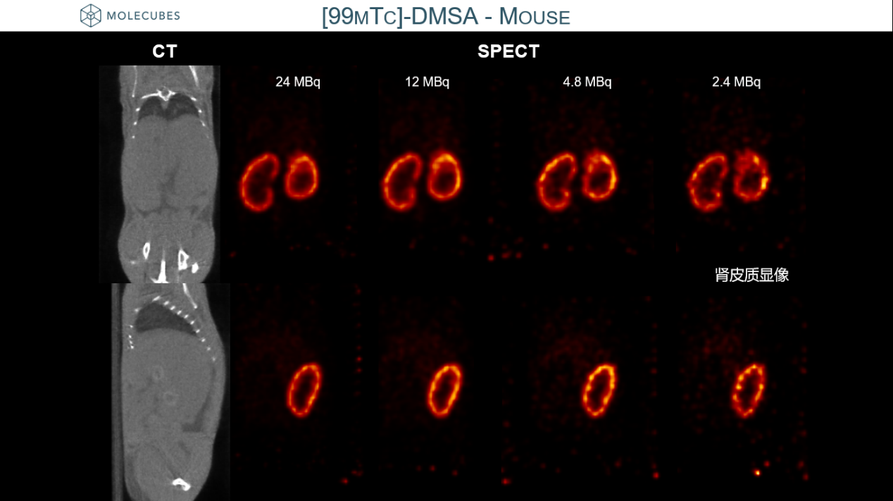
Comparison of SPECT images of different radiation doses: Mouse kidney imaging, [99mTc]DMSA, only renal cortex imaging, indicating that low dose imaging is possible.
5、Thyroid SPECT imaging
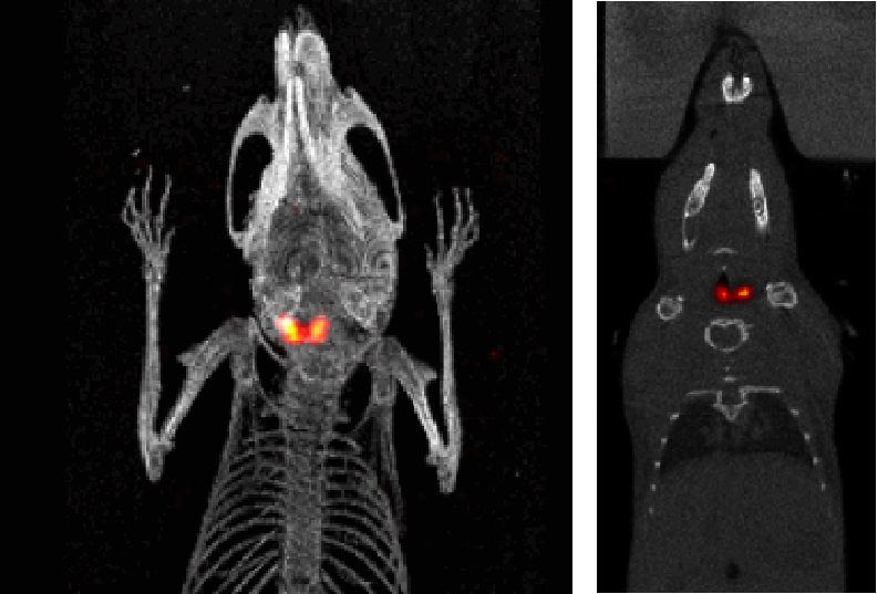
I123 Mouse thyroid SPECT imaging: The mouse thyroid was detected with 9.4MBq (250µCi) I123, and the acquisition time was 30min.
