Bruker BioSpec Preclinical MRI Instruments
Versatility
UltraShielded and Refrigerated
Maximum Space
Detail
Product Principles
Preclinical MRI enables a multitude of facets of basic research, disease, and treatment studies to be illuminated. Non-invasive in vivo imaging of small animals and rodents provides structural and functional information with high spatial and temporal resolution.
Brief Introduction
Leading scientists worldwide are taking their research closer to the molecular and cellular level thanks to BioSpec®. Designed for the emerging market of preclinical imaging and molecular MRI, its innovative modular concept enables virtually any small animal MR imaging application in life science, biomedical, and preclinical research.
BioSpec® already brings proven performance, safety, convenience, and future-proof cost-effective operation to over 600 labs worldwide. With our new multimodal animal platforms and accessories and our latest generation ParaVision® software more users than ever can access a new level of research quicker than they think.
Key Benefits
Cost effective - Helium zero boil off and Nitrogen-free for reduced maintenance costs and longer service intervals
Scalable – upgradeable electronics, options, and accessories to keep your system at the cutting edge
Compatible – multimodal animal handling solutions for use with all Bruker preclinical modalities
Intuitive – with Bruker’s gold standard preclinical imaging software, ParaVision®, users can start scanning immediately
Proven – more than 30 methods with various contrast modules and hundreds of measurement protocols designed and tested on mice and rats to guarantee the highest image quality
Key Technologies
Field strengths – 4.7 to 15.2 Tesla ultra-shielded supercon ducting magnets with reduced stray field
Bore sizes – 11 to 40 cm for optimal working space
Gradient diameters – 6, 9, 12, 20, 26 cm, with 6 and 12 cm gradients available as inserts (dual gradients)
Gradient strength – up to 1000 mT/m for highest duty cycles
Shim sets – Full 3rd order (6 cm) and 2nd order (9-26 cm) for best homogeneity
Product Characteristics
Zero helium boil-off technology, nitrogen-free, Ultra Shielded and Refrigerated (USR) magnet technology for reduced maintenance costs and longer service intervals
Accurate animal positioning with the motorized animal handling system for increased throughput
Full second order and z3 high power shim channels guarantee optimal performance for spectroscopy and MRI
Scalable AVANCE NEO electronics enabling both MRI and MRS applications and incorporating up to 16 receiver channels
Real-time spectrometer control for optimization of acquisition parameters during scan
High performance BGA-S-HP gradients with highest amplitudes and slew rates, shim strengths and duty cycles
Fast and easily mountable gradient/shim inserts for optimum gradient/shim performance for small samples available
Complete RF coil portfolio for mice and rats available, including coils for head, brain, cardiac, body, optogenetics, Arterial Spin Labeling, multi-channel array coils with up to 16 channels, and x-nuclei
MRI CryoProbes with 2 element, 4 element, or as 13C for mice as well as 4 element for rat delivering an exceptional increase in sensitivity
Intuitive ParaVision software for multi-dimensional MRI/MRS data acquisition, reconstruction, analysis and visualization including IntraGate based methods, UTE, and ZTE
In-house development and production of all key components (software, magnet, gradient, spectrometer, RF-coils) ensures the best performance and short repair times
Application Fields
With the power to conduct limitless imaging applications, BioSpec®opens up a whole new world of applications. Customers across the world undertake daily investigations into:
Angiography - flow contrast and flow analysis of the velocity of each voxel for glioblastoma and aneurysm studies
Cardiology - investigation of cardiac strain, ejection fraction, and septum defects, using triggered sequences or Bruker’s patented IntraGate for navigator based retrospective gating
Diffusion - visualization of disturbed pathology, such as in multiple sclerosis, epilepsy, and stroke tumors
fMRI - insight into the brain‘s function
Molecular MRI - imaging at the cellular level
Perfusion - with and without CA for tumor- detection, -neoangio genesis, and -vascularization, and disruption of the blood brain barrier
Spectroscopy – quantification of metabolic disorders and long term changes in metabolic processes
Application
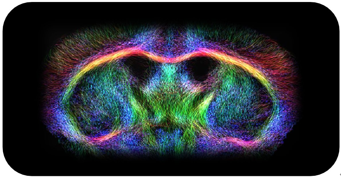
1、DTI Fibers
Brain connectivity studies in small animals are
challenging but can be achieved using state-ofthe-art MRI technologies for acquisition, such as the 7 T BioSpec® with MRI CryoProbe. Highresolution DTI MRI and fiber tracking of the living mouse brain delineate the organization of the
fiber tracts.
Acquisition details: MRI CyroProbeTM at 7 Tesla,
DTI-EPI, diffusion directions: 30, resolution:
(12.5 × 15.5 × 50) µm³, scan time: 25 min.

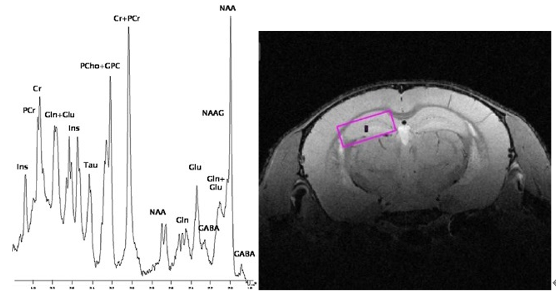
2、Localized Spectroscopy
STEAM spectrum of the mouse brain acquired using the MRI CryoProbeTM at 15.2 Tesla.
Acquisition details: STEAM, voxel size: (2 x 2 x 2) mm³, TR: 8 s, 128 averages, resolution enhancement with shifted Gauss filtering, TE: 1.1 ms, shift: 7%, broadening: 7 Hz

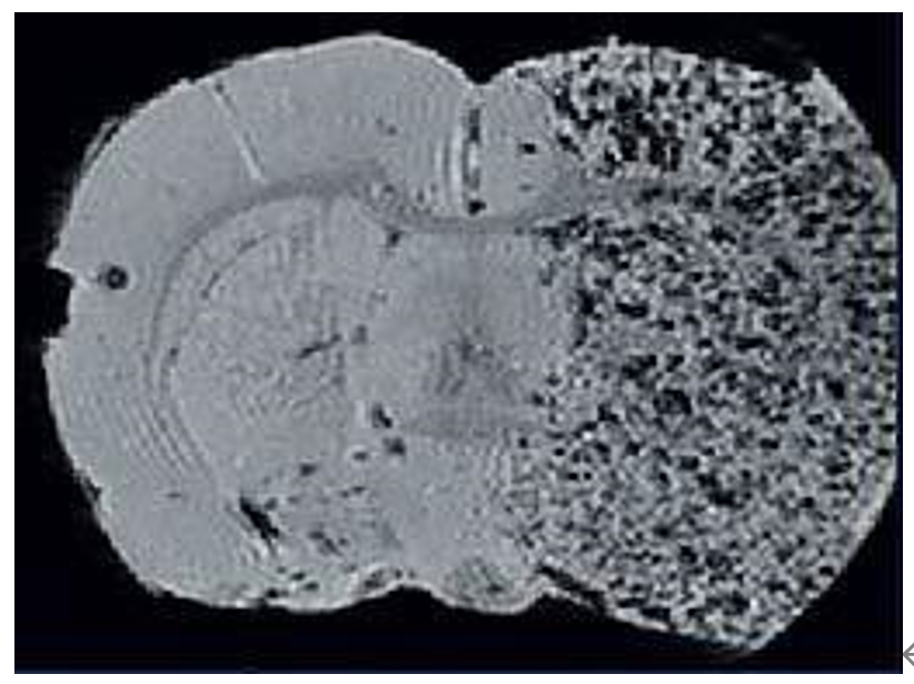
3、Cellular Imaging
Mesenchymal stem cells were labeled and used as markers for stroke regions. They are readily visible in T2- and T2*-weighted images both in vivo and ex vivo. Full 3D brain coverage enables quantification and volume rendering.


